Beta-amyloid plaques are abnormal protein deposits that are commonly associated with Alzheimer's disease and other neurodegenerative disorders. The amyloid PET is a crucial tool in the in-vivo assessment of these deposits and its evaluation has proven to strongly benefit from quantification.
Quantitative analysis is essential for monitoring disease progression over time, assessing treatment responses, and differentiating between various neurodegenerative disorders. DORIAN’s solution to amyloid PET quantification integrates traditional and innovative independent algorithms, overcoming the inherent limitations of each method to achieve more accurate results. This solution is validated alongside our clinical partners for all commercial tracers and includes two innovative patented algorithms:
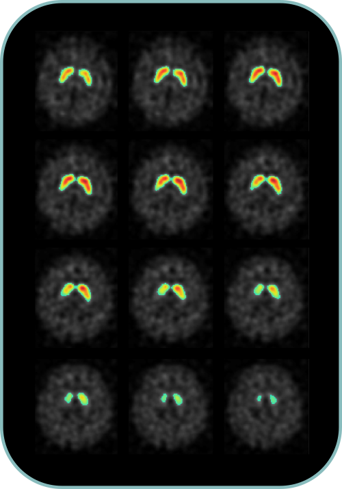
The 123I-Ioflupane SPECT images - commercially known as DaTSCAN or STRIASCAN -, are commonly used in the diagnostic pathway of movement disorders. To help the diagnostic evaluation, we developed a fully-automated software for computing a number of intensity or shape-based markers such as:
For each marker of each lobe, the marker value will be returned along with an age-corrected percentile value. An overall, machine-learning based probabilistic assessment will also be displayed to suggest a possible prediction of evaluation, based on more than a thousand manual gravity assessments run by consensus. This will produce an automated and data-driven report. The whole process is intended to facilitate the clinical report of a 123I-Ioflupane SPECT, especially in doubtful cases.
18F-fluoro-deoxy-glucose Positron Emission Tomography (FDG-PET) allows early identification of neurodegeneration in dementia. The use of an optimized method based on the SPM software package highly improves diagnostic accuracy.
Medial temporal lobe (MTL) atrophy from structural MR imaging is one of the key biomarkers to detect early neurodegenerative changes in the course of dementia and particularly Alzheimer's disease (AD). DORIAN's solution to 3D T1-weighted MRI quantification relies on an automated analysis pipeline build to assess the atrophy's progession in highly localized ROI of MTL producing a classification index with high accuracy that can also be used to predict the probability of AD conversion within a time frame of two years.